Mushroom Spores for Microscopy & Study
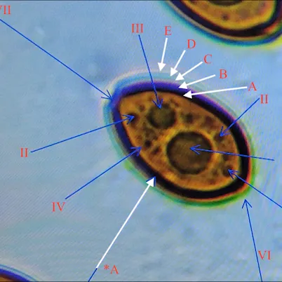
Mushroom spores are microscopic reproductive cells produced by fungi and are among the most useful structures for studying fungal morphology and taxonomy. Differences in spore size, shape, color, surface texture, and other microscopic characteristics can help researchers compare specimens and distinguish between species.
This guide explores the fundamentals of mushroom spores, how they are observed under a microscope, and the different collection formats used for microscopy, taxonomy, and long-term reference.
What Are Mushroom Spores?
Mushroom spores are microscopic reproductive cells produced by fungi as part of their reproductive life cycle. Although they are sometimes compared with plant seeds, fungal spores are biologically very different and are generally far smaller and structurally simpler.
In many mushroom-forming fungi, spores develop on specialized microscopic structures located on the fertile surface of the fruiting body. Once mature, they are released into the surrounding environment and may be carried by air currents, water, animals, or physical contact.
Spores can vary considerably between fungal species.
Mycologists examine characteristics such as:
– Overall shape
– Length and width
– wall thickness
– surface ornamentation
– pigmentation
– germ pore structure
reactions to particular microscopy stains or reagents
These characteristics can provide valuable taxonomic information, especially when microscopic observations are combined with macroscopic features such as cap shape, gill attachment, habitat, and spore-print color.
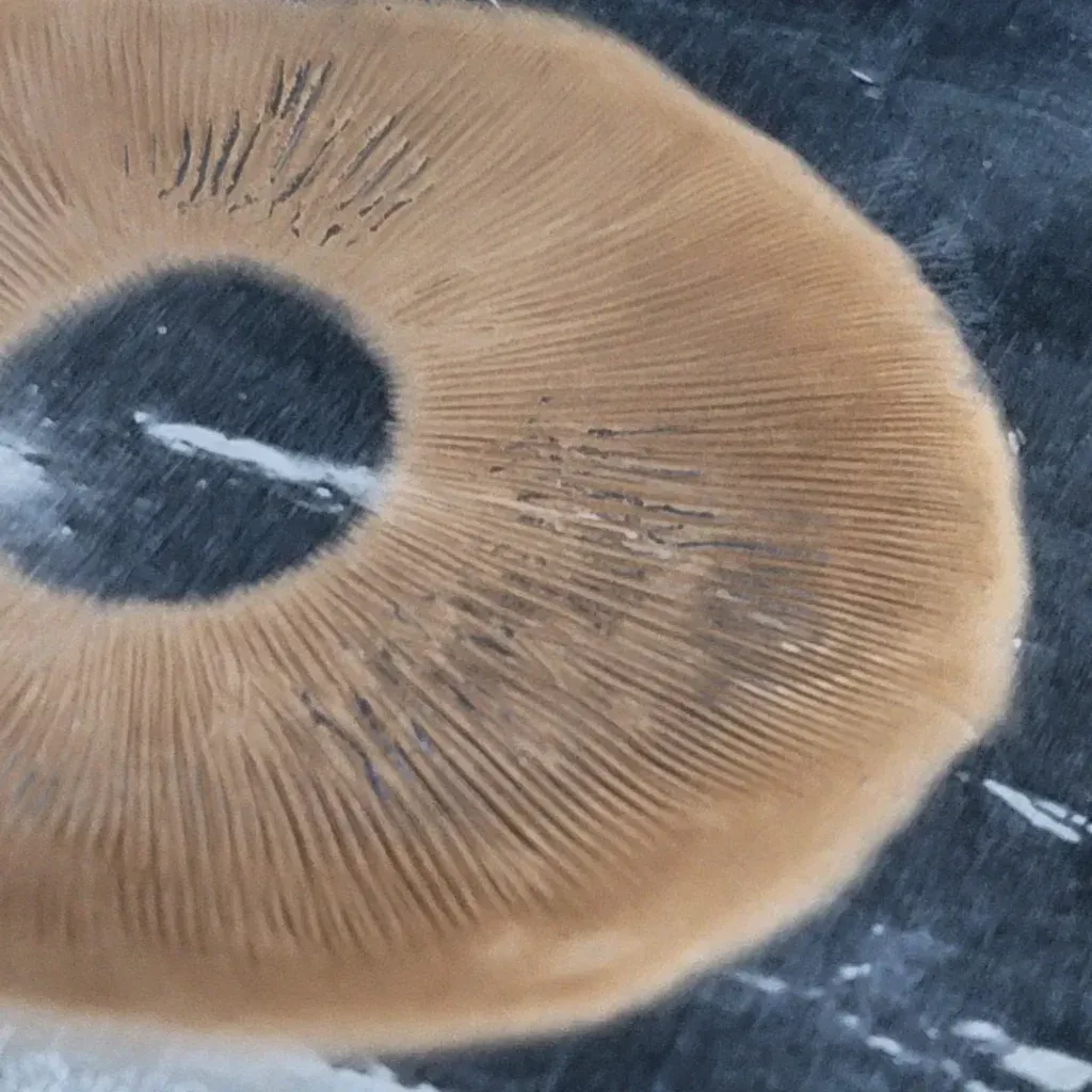
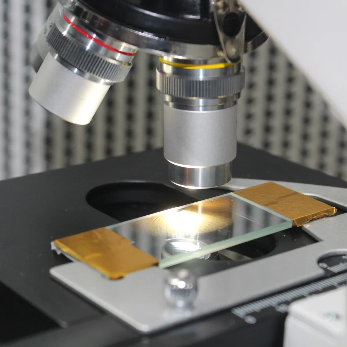
Studying Mushroom Spores Under a Microscope
Microscopy allows features of mushroom spores that are invisible to the naked eye to be examined directly. Depending on the species and magnification used, researchers may observe differences in spore shape, dimensions, pigmentation, wall structure, surface texture, and other identifying characteristics.
Spore measurements are particularly useful in fungal taxonomy. Rather than measuring a single spore, researchers typically examine multiple spores from the same collection to understand the natural range of variation within a specimen.
Microscopy can also reveal structures associated with spores and the surrounding fungal tissue, helping researchers build a more complete picture of a specimen’s morphology.
Foil Spore Prints
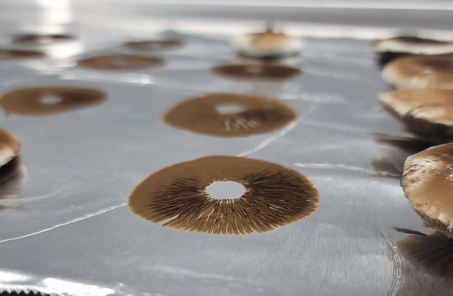
A spore print is created when mature spores released from a mushroom accumulate on a surface beneath the cap. The resulting deposit can reveal the overall color of the spores while preserving material that may later be examined under a microscope.
Foil is commonly used because it is lightweight, easy to store, and provides a clean surface from which small samples can be collected for observation.
Spore prints are particularly useful for:
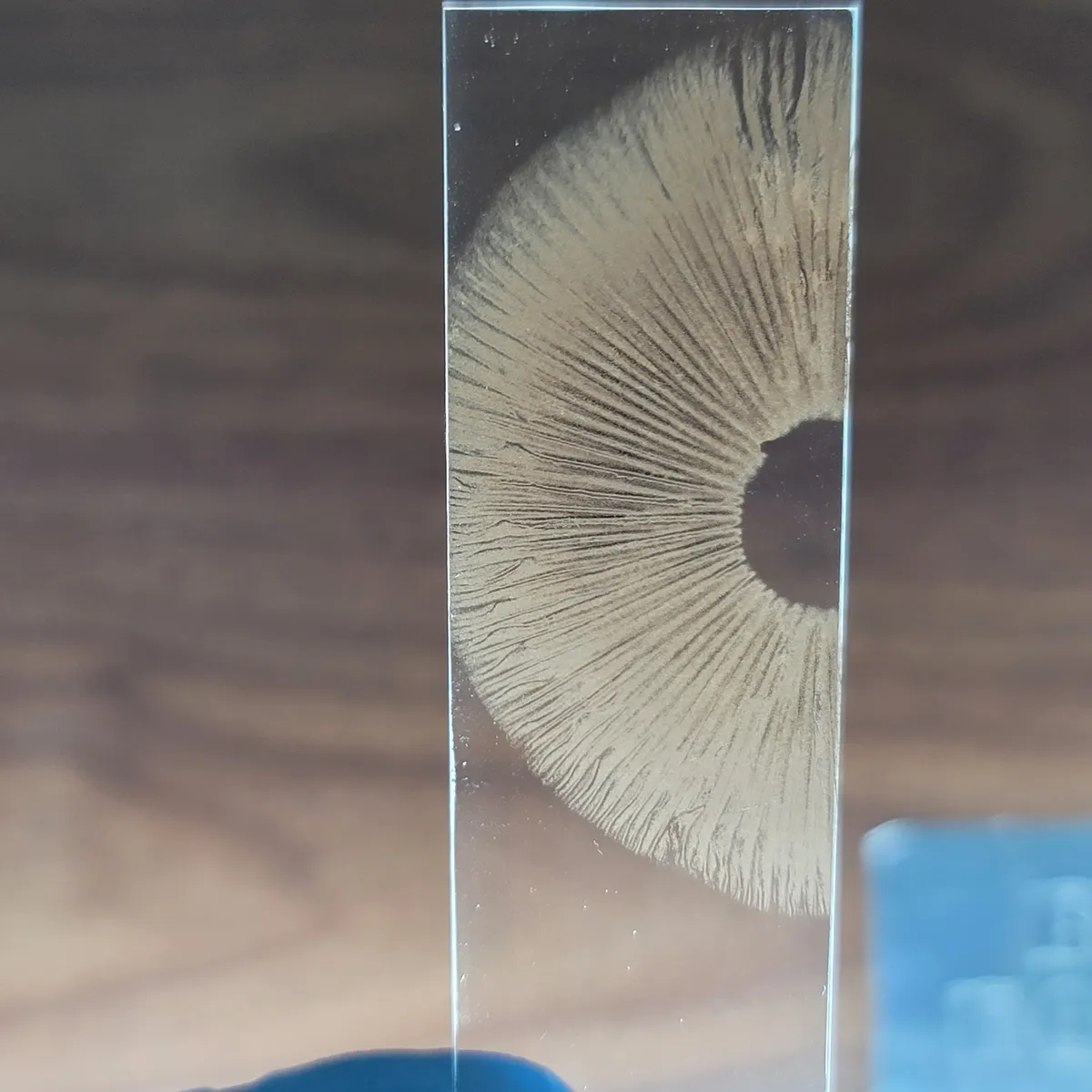
Microscopy — Small portions of the deposit can be transferred to a slide for examination.
Spore Color Reference — The accumulated deposit can reveal colors that may be difficult to see when examining individual spores.
Taxonomic Comparison — Prints can be preserved alongside photographs, collection notes, and microscopy observations.
Long-Term Collections — Properly stored prints take up very little space and can serve as useful reference material for future study.
Featured Spore Prints
-
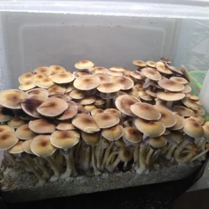
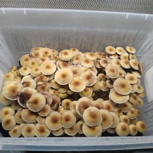
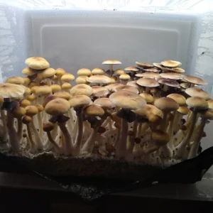
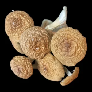
 Product on saleBAPER RUST Spore PrintOriginal price was: $30.00.$20.00Current price is: $20.00.
 -
Product on saleBlue Meanie Spore PrintOriginal price was: $30.00.$20.00Current price is: $20.00.
-
Product on saleGolden Teacher Spore PrintOriginal price was: $30.00.$20.00Current price is: $20.00.
Spore Syringes
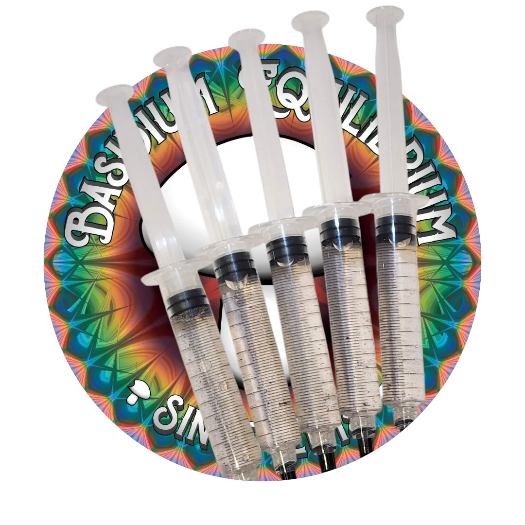
A spore syringe contains fungal spores suspended in a liquid solution. Because spores are dispersed throughout the solution, a small sample can be placed onto a microscope slide for observation without removing material from a dry spore print.
Under magnification, researchers can examine individual spores and compare characteristics such as size, shape, pigmentation, and structural variation.
Spore syringes also provide a convenient way to keep a microscopic sample contained and labeled as part of a reference collection.
Why Use Spore Syringes for Microscopy?
Controlled Sampling — A small amount of solution can be transferred directly to a microscope slide.
Contained Storage — Samples remain enclosed until they are needed for observation.
Repeat Observation — Multiple slides can be prepared from the same reference sample.
Microscopic Comparison — Individual spores can be examined alongside samples from other collections or species.
Spore Swab
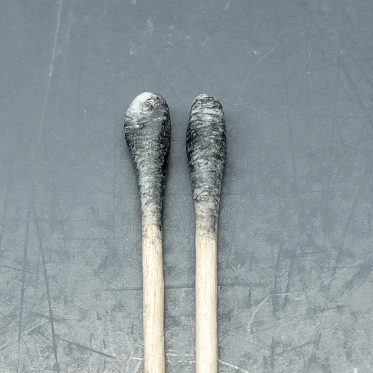
Spore swabs provide another method for collecting microscopic fungal material. A sterile swab can be passed across a spore-bearing surface, allowing spores to adhere to its fibers for later observation.
Swabs are particularly useful when a specimen produces only a faint spore deposit or when obtaining a traditional spore print is difficult.
For collectors and researchers, swabs can provide a compact way to preserve material associated with a documented specimen.
Why Are Spore Swabs Useful?
Direct Collection — Spores can be collected directly from a specimen.
Useful for Sparse Deposits — Helpful when a clearly visible print is difficult to obtain.
Compact Storage — Swabs require very little storage space.
Microscopy Reference — Material can be transferred from the swab to a microscope slide for examination.
References & Further Reading
A selection of foundational texts and research sources for those interested in mushroom spores, taxonomy, and fungal biology. These references have shaped modern understanding of fungal taxonomy and microscopy.
Gastón Guzmán
The Genus Psilocybe: A Systematic Revision of the Known Species
A foundational work in fungal taxonomy, detailing spore morphology, classification, and species distribution.
Nicholas P. Money
Mushroom — Oxford University Press
An accessible overview of fungal biology, including spore structure, dispersal, and development.
Paul Stamets
Mycelium Running: How Mushrooms Can Help Save the World
Cornell University
Buller’s Drop and Spore Discharge Mechanics
A quantitative study examining how mushroom structure influences spore release and distribution.
sciencedirect.com.com
Spore Science Made Simple
Curious about spores? These are the six most common questions beginners ask, answered in clear and simple detail to help you build a strong foundation in spore science.