Psilocybe Tampanensis
The mushroom behind the legendary philosopher’s stones – tampanensis is famous for producing underground fungal structures more widely known as magic truffles.
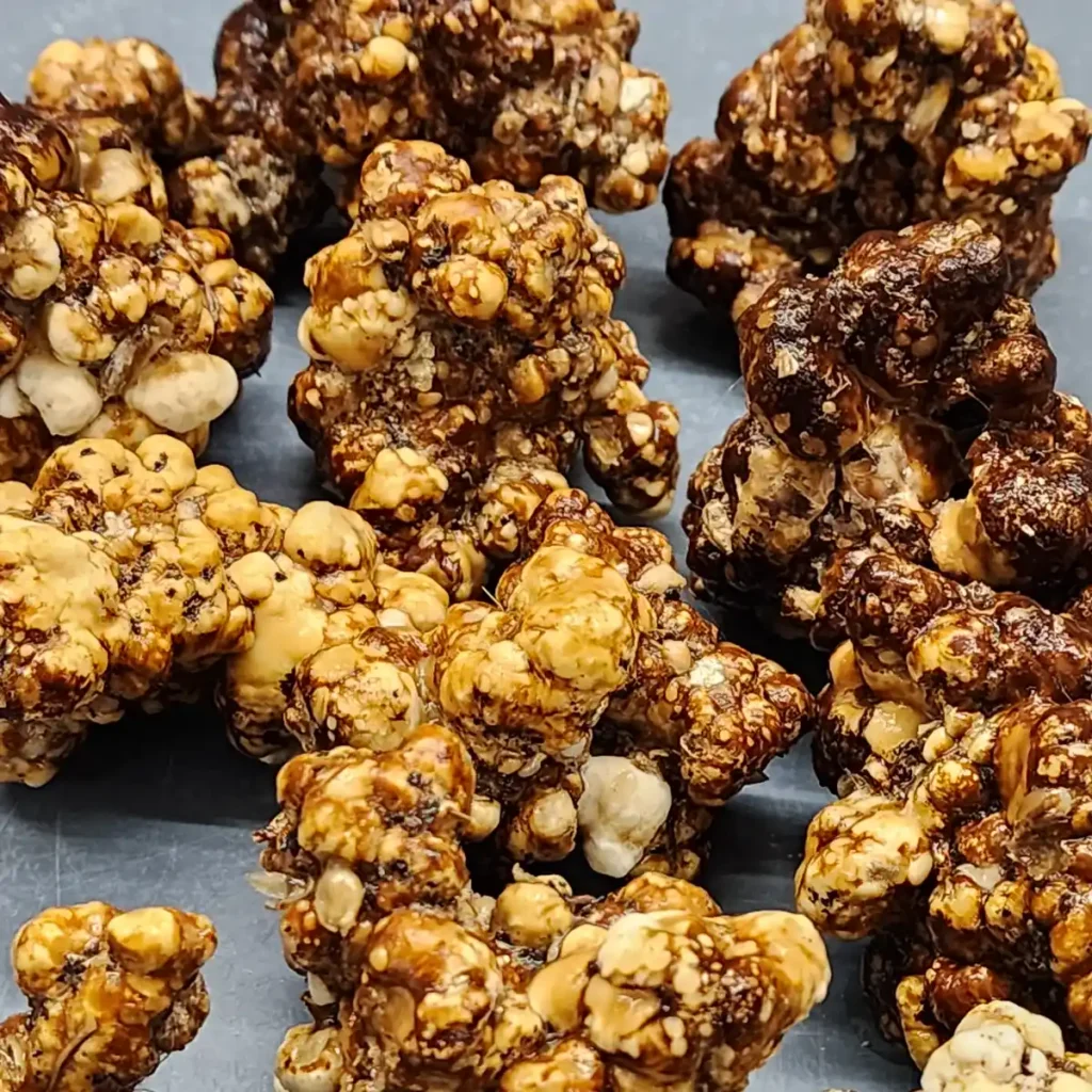
Wild Mississippi Tampanensis
Few mushrooms have built a reputation quite like Psilocybe tampanensis. Often associated with the underground fungal structures known as philosopher’s stones — or more commonly magic truffles — this species became widely known through its presence in Amsterdam, where sclerotia have long been legally sold and studied. Unlike typical mushrooms that are valued primarily for their fruiting bodies, tampanensis is famous for producing dense subterranean sclerotia, a survival structure that has fascinated mycologists and enthusiasts for decades.
Over time, a small number of laboratory-maintained lineages came to dominate most observations of this species. Among them, the well-known ATL7 isolate became a reference point for tampanensis genetics due to its reliable formation of large, dense sclerotia.
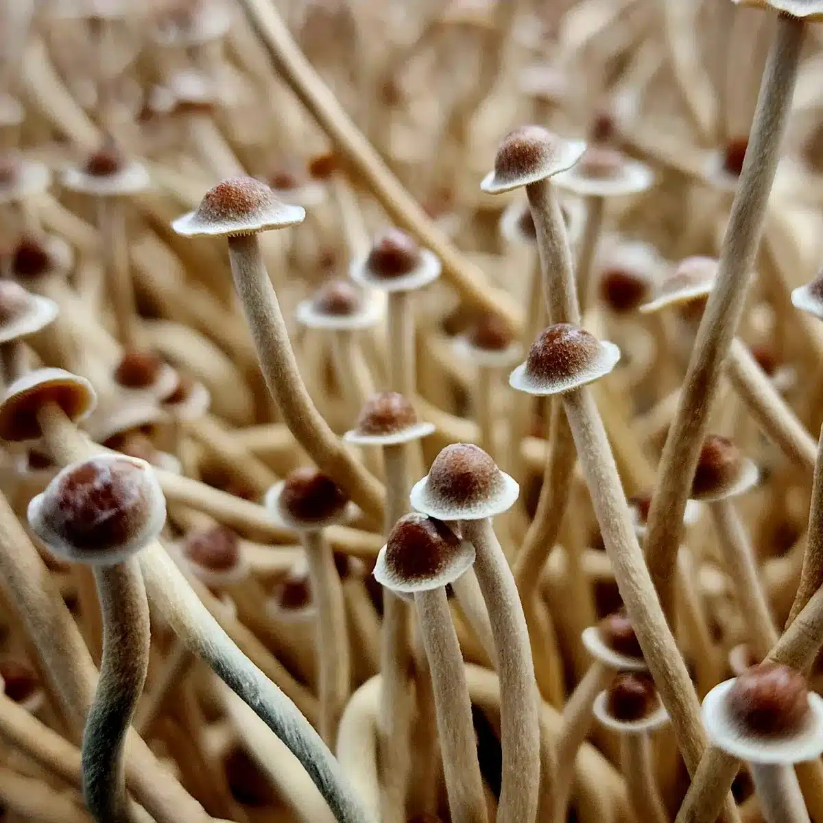
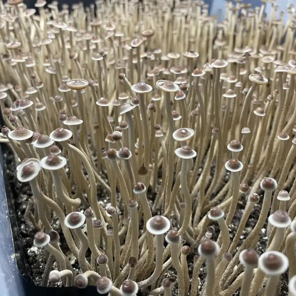
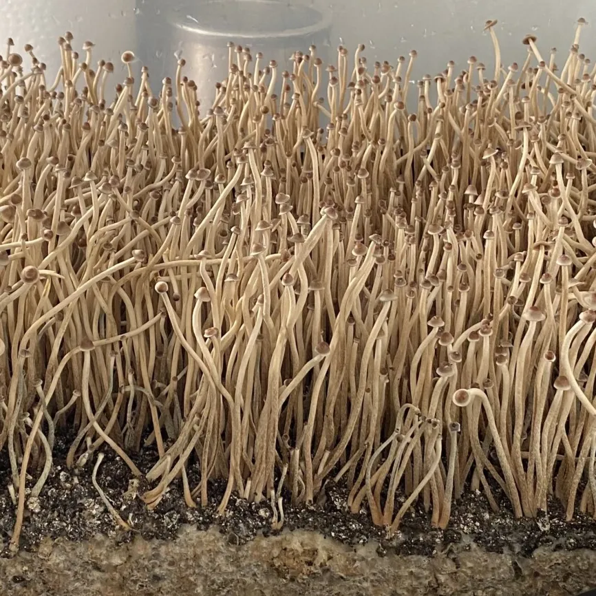
More recently, however, new genetic material has surfaced from the southeastern United States. This wild Mississippi strain of Psilocybe tampanensis was discovered by a citizen scientist and later sequenced and cataloged, offering a rare look at a lineage that has not been heavily domesticated through decades of laboratory propagation. Compared with established lines like ATL7, this wild variant displays smaller fruiting bodies with slender stems and compact caps while still producing solid sclerotia.
The Mississippi lineage is currently maintained as an exclusive genetic line within the Basidium Equilibrium collection, representing a unique opportunity to observe the natural diversity that exists within the tampanensis species.
What Are Philosopher’s Stones?
Unlike most mushrooms that reproduce primarily through visible fruiting bodies, Psilocybe tampanensis is famous for forming dense underground structures known as sclerotia. These compact masses of mycelium act as a survival mechanism for the fungus, allowing it to store nutrients and endure unfavorable environmental conditions.
Over time, these hardened mycelial formations became popularly known as “philosopher’s stones,” a name inspired by alchemical lore and the mysterious appearance of the structures themselves. In modern culture they are also commonly referred to as magic truffles, particularly in the Netherlands where several sclerotia-producing psilocybin species gained widespread recognition through the retail truffle market in Amsterdam.
From a biological perspective, philosopher’s stones are not mushrooms in the traditional sense. They are instead a dense aggregation of fungal tissue, formed underground as the mycelium consolidates nutrients into small, firm nodules. When conditions become favorable, the fungus may eventually produce surface fruiting bodies, but the sclerotia themselves serve as a resilient life stage within the organism’s lifecycle.
Because of their unusual structure and role in fungal survival, sclerotia produced by Psilocybe tampanensis have attracted significant attention among mycologists studying fungal adaptation, reproductive strategies, and genetic variation within the genus.
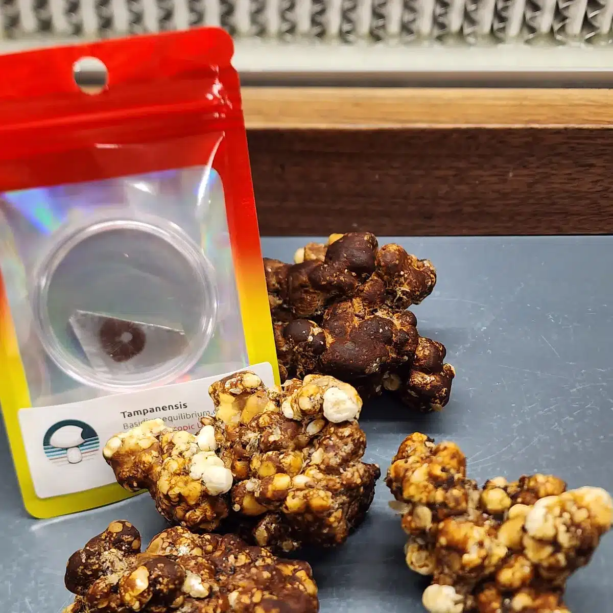
Microscopy Characteristics of Psilocybe tampanensis
Under magnification, Psilocybe tampanensis reveals many of the defining microscopic traits associated with the genus Psilocybe. Careful observation of spores, basidia, and cystidia provides valuable insight into species identification and taxonomy, making microscopy an essential tool for studying tampanensis and closely related species.
The spores of Psilocybe tampanensis are typically ellipsoid to sub-ellipsoid in shape and display the characteristic dark purplish-brown coloration common among psilocybin-producing species within the genus. When viewed under high magnification, the spores show a smooth surface and a distinct germ pore, a small opening that plays a role in the early stages of spore germination.
Basidia — the spore-producing cells located on the gills — are generally four-spored, though variation can occasionally occur. These microscopic structures are responsible for producing and releasing spores that allow the fungus to reproduce and disperse.
Additional microscopic features may include the presence of cheilocystidia along the gill edges, which can vary in shape from flask-like to lageniform. These structures, along with spore morphology, help distinguish tampanensis from other closely related Psilocybe species during microscopic examination.
For researchers and microscopy enthusiasts, examining tampanensis spores provides an opportunity to observe the intricate structures that define fungal reproduction. Combined with macroscopic observations of fruiting bodies and sclerotia formation, these microscopic characteristics help build a clearer understanding of the species and its place within the broader Psilocybe lineage.